Αρχειοθήκη ιστολογίου

Αναζήτηση αυτού του ιστολογίου

Κυριακή 6 Δεκεμβρίου 2020

Madhura mycosis of foot

xlomafota13 shared this article with you from Inoreader


MADHURA%2BFOOT%2BMRI.jpg

Sagittal T2w
MADHURA%2BFOOT%2BRING%2BSIGN.jpg

Sagittal T1w
MADHURAMYCOSIS%2BFOOT%2BMRI.jpg


Sagittal STIR

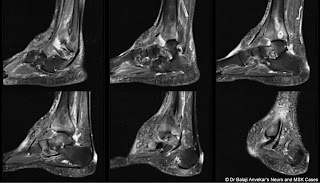
MRI study of ankle/foot with x-ray correlation shows:

A lobulated abnormal soft tissue measuring approximately 70 mm in length and 40 mm in depth on dorsal aspect of foot encasing extensor tendons with hypo intense signal on T2-weighted images, "dot in a circle" sign on MRI. 

Soft tissue density on x-ray without dystrophic calcification on x-ray. Lytic destruction of adjacent anterior corner of tibia on MRI and x-ray. Associated tibio talar joint effusion. Multifocal ovoid l ytic lesion with sclerotic rim on x-ray involving distal end of tibia with fluid signal on MRI. Marginal lytic destruction of distal end of fibula. Circumferential punched-out marginal erosion of neck of talus which is markedly thinned out with an associated marrow oedema on STIR. Multifocal marrow oedema involving tarsal bones, tenosynovitis of extensor as well as plantar tendons.

Multiple ulcers, nodules and discharging sinuses on skin of dorsal aspect of foot when examined clinically.

Imaging diagnosis: Madhura mycosis of foot with osteomyelitis of tibia.

View on the web

COVID 19 Cerebritis

xlomafota13 shared this article with you from Inoreader

Clinically: A known case of COVID 19 positive admitted for fever and breathlessness.

After five days of hospital admission developed sinusitis, headache and started worsening repidly. Subjected for MRI due to sudden onset loss of consciousness and neurological examination revealed new onset ophthalmoplegia. 

COVID%2BCEREBRITIS%2BMRI%2BBRAIN.jpg

COVID%2BCEREB   RITIS%2BPNS%2BSPREAD.jpg

COVID%2BCEREBRITIS.jpg

CORONA%2BBRAIN%2BINFECTION.jpg

COVID%2BBRAIN%2BINFECTION%2BDW.jpg


MRI shows bilateral geographic shaped patchy T2 hyperintensities involving frontal lobes with diffusion restriction at the floor of anterior cranial fossa. Mild lepto meningeal enhancement on post contrast. 
An associated marked bilateral paranasal sinusitis.

Possibility of COVID 19 Cerebritis, Neuro inv asiveness by transnasal route suggested and can be attributed to known Neurotropism of the virus. 

Neurotropism of Covid 19

Neuronal pathway is one of important way of spread of neuropathic viruses like Cov to enter central nervous system. These viruses can migrate with the help of sensory as well as motor nerve endings and have ability of retrograde as well as antegrade spread along the olfactory nervous system due to the unique anatomical organisation of olfactory nerves and olfactory bulb in the nasal cavity and fore brain. As a result, Cov after paranasal sinus infection can enter brain through olfactory tract in early stages of infection rapidly, within seven days of infection as in our case.


View on the web

Reconstruction of upper limb soft-tissue defects after sarcoma resection with free flaps: A systematic review.

xlomafota13 shared this article with you from Inoreader
Related Articles

Reconstruction of upper limb soft-tissue defects after sarcoma resection with free flaps: A systematic review.

J Plast Reconstr Aesthet Surg. 2020 Nov 08;:

Authors: Lucattelli E, Lusetti IL, Cipriani F, Innocenti A, De Santis G, Innocenti M

Abstract
BACKGROUND AND OBJECTIVES: Upper limb preservation after soft tissue sarcoma (STS) surgical excision is now the accepted gold standard and it often requires reconstruction with free flaps. The purpose of this review is to summarize current literature on upper limb reconstruction with free flaps after STS resection.
METHODS: A systematic review was performed in July 2019 in PubMed and MedLine Ovid databases according to the PRISMA guidelines.
RESULTS: A total of 17 studies were included in the final analysis, with 132 patients. The most common diagnosis was Malignant Fibrous Histiocytoma. The most frequent timing of flap coverage was immediate. The success rate was almost always 100%. The length of follow-up was reported in 11 studies with a range of 2-187 months. The most commonly reported patient-centered outcome was the MSTS Score. Based on the evidence of the literature collected, we divided the upper limb into four parts (shoulder, elbow and arm, forearm and wrist, and hand) and described the most common and functional free flaps used for reconstruction after STS resection.
CONCLUSIONS: Free flaps in the treatment of STS of the upper extremity have a good overall outcome, with a low postoperative complication rate. A wide array of free flaps is available for reconstruction, and the choice of flap is based on defect size, types of tissue required, postoperative functional goal, and surgeon preference. A greater degree of standardization is needed in the reporting of patient-centered outcomes to facilitate future comparative studies.

PMID: 33277214 [PubMed - as supplied by publisher]

View on the web

Long-term functional outcomes of vascularized fibular and iliac flap for mandibular reconstruction: A systematic review and meta-analysis.

xlomafota13 shared this article with you from Inoreader
Related Articles

Long-term functional outcomes of vascularized fibular and iliac flap for mandibular reconstruction: A systematic review and meta-analysis.

J Plast Reconstr Aesthet Surg. 2020 Nov 08;:

Authors: Ma H, Van Dessel J, Shujaat S, Bila M, Gu Y, Sun Y, Politis C, Jacobs R

Abstract
INTRODUCTION: To date, there is a lack of evidence related to the long-term evaluation of recipient-site functional outcomes following mandibular reconstruction with vascularized bone grafts. Therefore, the aim of this systematic review and meta-analysis was to evaluate the long-term recipient-site functional outcomes in oral oncology patients who require mandibular reconstruction with either vascularized fibular flap (VFF) or vascularized iliac flap (VIF).
METHODS: An extensive electronic search was conducted in PubMed, Web of Science, Cochrane, and Embase databases for identifying articles published until April 2020. All papers were dual screened for eligibility in accordance with the Preferred Reporting of Systematic Reviews and Meta-Analyses and Meta-analysis of Observational Studies in Epidemiology guidelines. The risk of bias was assessed using the MINORS tool. A meta-analysis of functional outcome parameters was performed to estimate single incidence rates.
RESULTS: A total of 257 patients with a mean follow-up period of 38.6 ± 19.5 months were included in this meta-analysis, where 174 patients underwent VFF reconstruction and 83 patients involved reconstruction with VIF. The functional outcomes in patients reconstructed with VIF showed improved scoring for mastication, deglutition, diet, and speech. Speech showed highest score among all functional parameters, whereas, mastication was the most poorly recovered parameter in relation to reconstruction with both flaps. No significant difference in functional outcomes was observed between both flaps.
CONCLUSION: Current evidence seems to indicate that VIF offers improved long-term recipient-site functional outcomes. Yet, considering a high level of data heterogeneity in published studies, future long-term standardized comparative studies should be conducted.

PMID: 33277215 [PubMed - as supplied by publisher]

View on the web

Magnetic resonance lymphography as three-dimensional navigation for lymphaticovenular anastomosis in patients with leg lymphedema.

xlomafota13 shared this article with you from Inoreader
Related Articles

Magnetic resonance lymphography as three-dimensional navigation for lymphaticovenular anastomosis in patients with leg lymphedema.

J Plast Reconstr Aesthet Surg. 2020 Nov 08;:

Authors: Yasunaga Y, Nakajima Y, Mimura S, Yuzuriha S, Kondoh S

Abstract
BACKGROUND: Precise mapping of functional lymphatic vessels is essential for successful lymphaticovenular anastomosis (LVA). This study aimed to clarify the precision of magnetic resonance lymphography (MRL) in detecting lymphatic vessels prior to LVA.
METHODS: Eighteen patients with leg lymphedema were recruited for this prospective study. All patients underwent MRL before LVA to obtain three-dimensional coordinates of lymphatic vessels from MRL images. The precision of MRL for detecting lymphatic vessels was evaluated and compared with those of other contrast techniques.
RESULTS: Twenty legs from 18 patients were analyzed. A total of 40 skin incisions were made, 32 of which were determined by MRL. The precision of MRL to detect lymphatic vessels was 94%. With the addition of MRL, the number of lymphatic vessels identified preoperatively was increased as compared with indocyanine green lymphography (ICG-L) alone. Assuming a detection sensitivity of MRL for lymphatic vessels of 1, those of other contrast techniques were 0.90 for ICG-L under microscopy, 0.73 for patent blue staining, and 0.43 for ICG-L before incision. Whereas ICG-L before incision could not detect lymphatic vessels at depths greater than 17.0 mm, all deeper anastomosed lymphatic vessels were identified by MRL.
CONCLUSION: Lymphatic vessels enhanced on MRL can be reliably identified intraoperatively. MRL is a promising preoperative examination in LVA that can selectively depict suitable lymphatic vessels even in deep tissue layers.

PMID: 33277216 [PubMed - as supplied by publisher]

View on the web

Πέμπτη 3 Δεκεμβρίου 2020

Eosinophilic Panniculitis Associated With COVID-19

xloma.fota.13 shared this article with you from Inoreader
V.M. Leis-Dosil, A. Sáez Vicente, M.M. Lorido-Cortés
Actas Dermosifiliogr. 2020;111:804-5

Full Text - PDF
View on the web

Managing Psoriasis Consultations During the COVID-19 Pandemic: Recommendations From the Psoriasis Group of the Spanish Academy of Dermatology and Venereology (AEDV)

xloma.fota.13 shared this article with you from Inoreader
I. Belinchón, L. Puig, L. Ferrándiz, P. de la Cueva, J.M. Carrascosa
Actas Dermosifiliogr. 2020;111:802-4

Full Text - PDF
View on the web