Αρχειοθήκη ιστολογίου

Αναζήτηση αυτού του ιστολογίου

Πέμπτη 28 Ιανουαρίου 2021

Auswirkungen der SARS-CoV‑2-Pandemie auf die universitäre Hals-Nasen-Ohren-Heilkunde im Bereich der Forschung, Lehre und Weiterbildung

Greek DailyNews shared this article with you from Inoreader
Via HNO

106.jpg

Zusammenfassung

Hintergrund

Ab Frühjahr 2020 kam es zur weltweiten Verbreitung von SARS-CoV‑2 mit der heute als erste Welle der Pandemie bezeichneten Phase ab März 2020. Diese resultierte an vielen Kliniken in Umstrukturierungen und Ressourcenverschiebungen. Ziel unserer Arbeit war die Erfassung der Auswirkungen der Pandemie auf die universitäre Hals-Nasen-Ohren(HNO)-Heilkunde für die Forschung, Lehre und Weiterbildung.

Material und Methoden

Die Direktorinnen und Direktoren der 39 Universitäts-HNO-Kliniken in Deutschland wurden mithilfe einer strukturierten Online-Befragung zu den Auswirkungen der Pandemie im Zeitraum von März bis April 2020 auf die Forschung, Lehre und die Weiterbildung befragt.

Ergebnisse

Alle 39 Direktorinnen und Direktoren beteiligten sich an der Umfrage. Hiervon gaben 74,4 % (29/39) an, dass es zu einer Verschlechterung ihrer Forschungstätigkeit infolge der Pandemie gekommen sei. Von 61,5 % (24/39) wurde berichtet, dass pandemiebezogene Forschungsaspekte aufgegriffen wurden. Von allen Kliniken wurde eine Einschränkung der Präsenzlehre berichtet und 97,5 % (38/39) führten neue digitale Lehrformate ein. Im Beobachtungszeitraum sahen 74,4 % der Klinikdirektoren die Weiterbildung der Assistenten nicht gefährdet.

Schlussfolgerung

Die Ergebnisse geben einen Einblick in die heterogenen Auswirkungen der Pandemie. Die kurzfristige Bearbeitung pandemiebezogener Forschungsthemen und die Einführung innovativer digitaler Konzepte für die studentische Lehre belegt eindrücklich das große innovative Potenzial und die schnelle Reaktionsfähigkeit der HNO-Universitätskliniken, um auch während der Pandemie ihre Aufgaben in der Forschung, Lehre und Weiterbildung bestmöglich zu erfüllen.

View on the web

Traumatic brain injury: 10 things you need to know to save lives

Greek DailyNews shared this article with you from Inoreader

Message:

1. THERE'S LITTLE ROOM TO SPARE IN THE CRANIUM
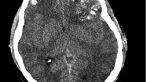
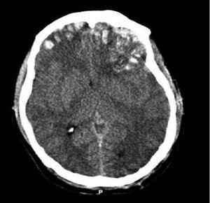
TBI can cause inflammation and swelling or bleeding from damage to brain tissue and blood vessels. Bleeding may occur within the brain (an intracranial hemorrhage) or the protective layers that surround it, such as an epidural, subdural or subarachnoid hemorrhage.

TBI can cause inflammation and swelling or bleeding from damage to brain tissue and blood vessels.
TBI can cause inflammation and swelling or bleeding from damage to brain tissue and blood vessels. (Photo courtesy of Rehman T, Ali R, Tawil I, Yonas H)
RELATED ARTICLES
Stroke: 10 things you need to know to save lives
STEMI: 10 things you need to know to save lives
Trauma: 10 things you need to know to save lives
The brain is tightly enclosed within the skull. Bleeding and inflammation cause a rise in intracranial pressure (ICP), which squeezes and damages brain structures, within that space. As ICP increases, sections of the brain may shift to different areas within the skull, or into the opening where the spinal cord enters the cranium, through a process known as herniation. The higher and longer ICP rises, the degree of permanent disability and likelihood of death increases.

Signs of increased ICP include altered mental status, unequal or nonreactive pupils, posturing, or seizures, as well as bradycardia and hypertension (Cushing's Phenomenon).

2. SLOW DOWN THE DAMAGE FROM SECONDARY BRAIN INJURY
Brain tissue does not regenerate, thus damage from the initial insult is generally permanent. The subsequent edema and bleeding from the primary injury then spreads to damage other areas of the brain, and treatment is aimed at limiting this secondary injury. Through airway protection, oxygenation or ventilation, blood pressure maintenance, and transport to a trauma center, EMS plays a vital role in stopping this cascade.

3. HEAD INJURED PATIENTS ARE UNRELIABLE HISTORIANS
Patients with TBI may be confused, combative, or unresponsive. They may be unable able to follow commands or report pain, and concurrent drug or alcohol intoxication makes assessment even more difficult. Assume that altered mentation is caused by a head injury instead of intoxication.

Rely on inspection, auscultation, and palpation to rule in other injuries, such as a change in the patient's facial expression when their abdomen is palpated. Remember that internal injuries cannot be ruled out, particularly those involving the spinal cord, so take spinal precautions in any patient with a head injury and altered mental status.

4. SOUND THE ALARM AT THE TRAUMA CENTER
Treatment for head-injured patients requires rapid assessment, imaging with a CT scan, and possibly neurosurgery. Time is brain, and the chance of disability or death increases with any delay.

Transporting patients to trauma centers that offer neurological services, and early activation of teams at these centers, streamlines the process to definitive care. Know which hospitals these are, initiate transport as quickly as possible, and alert the hospital to prepare staff and equipment for your arrival.

5. AIRWAY COMPROMISE FROM TBI COMES IN MANY FORMS
Unconscious head-injured patients may lose muscle tone in their jaw, and their tongue may obstruct the airway. The gag reflex may also be compromised in TBI patients, which increases the risk of aspiration from vomit or blood. Clenched teeth, known as trismus, is another common finding that makes position and suctioning airway secretions difficult. Attempt to open the airway with a jaw thrust. Use an oral or nasal airway if the patient will tolerate it, position the patient on their left side to help prevent aspiration, and suction secretions.

Endotracheal intubation is the definitive airway management for TBI, often after sedation and paralysis with RSI. This procedure carries considerable risk, and whether it should be done by ground EMS crews is controversial. Remember that the goal for airway management should be to use the least invasive means to maintain a clear path for oxygenation and ventilation.

6. HYPOXIA AND HYPOTENSION ARE HARMFUL – EVEN FOR A SHORT TIME
Irreversible brain damage can occur in TBI patients after only four minutes of anoxia, which can be caused by a compromised airway, altered respiratory patterns from the head injury, or lung injury in multi-system trauma. Use pulse-oximetry, skin color, and respiratory rate to assess adequate oxygenation. Administer oxygen via nasal cannula, non-rebreather mask, or bag valve to maintain a pulse-ox reading of at least 95%.

Cerebral oxygen delivery is also compromised by hypotension, which is associated with poor outcomes after even transient episodes. Hypotension caused directly by a head injury is a rare and ominous finding; it is more often caused by shock associated with other injuries. Administer IV fluids and titrate blood pressure to 90 – 100 mmHG systolic in head injured patients who are hypotensive.

7. VENTILATING TOO FAST REDUCES BLOOD FLOW TO THE BRAIN
As harmful as hypoxia is in TBI, hypocapnea from excessive ventilation is harmful as well. A higher respiratory rate decreases the amount of CO2 in the blood, which triggers cerebral vasoconstriction and less oxygen delivery. Hyperventilation of head injuries has been associated with poor outcomes.

When assisted ventilation is necessary in isolated head injuries, follow your local protocols regarding use of end-tidal CO2 (EtCO2) monitoring for a head-injured patient.

Shock from multisystem trauma can also cause a low EtCO2 reading, in which case it should not be used to guide ventilation. If capnography is not available, or if the patient may be in shock from other injuries, ventilate adults at 10 breaths per minute. There is one exception...

8. HYPERVENTILATION MAY SLOW HERNIATION
In cases of suspected ICP being so high that portions of the brain may be shifting out of the cranium, the benefit of temporarily reducing cerebral blood with mild hyperventilation may outweigh the harm from less oxygen delivery. Hyperventilation is ONLY indicated for patients who are posturing or have no motor response, have unequal or dilated and nonreactive pupils, or bradycardia and hypotension. Titrate the respiratory rate to maintain an EtCO2 reading between 30 and 35 (in the absence of multi-system trauma), or 20 breaths per minute.

9. YOU MAY HAVE A CONCUSSION
A concussion is a transient change in mentation after blunt head trauma. It can cause retrograde amnesia, and patients often repeatedly ask the same question after being given an answer. It can also cause a temporary loss of consciousness, disorientation, incoherent speech, or lack of coordination. Diagnosis of a concussion is made after a CT scan shows no structural abnormalities associated with these symptoms - not in the field.

While concussion symptoms typically resolve after a few hours or days, severe or multiple concussions can cause headaches, dizziness, depression, and difficulty concentrating for weeks. Especially worrisome is a secondary impact syndrome, where a patient sustains a second concussion before recovering from the first. This can cause rapid cerebral edema and herniation, and has a high mortality rate. Athletes with a head injury should not return to play until after symptoms have completely resolved.

10. BE ON THE LOOKOUT FOR BLOOD THINNERS
Patients taking blood thinners, including Coumadin and Plavix, are at risk of severe bleeding into the brain from relatively minor head injuries. Learn what medications these are, and factor the patient's medication list into your assessment. Signs and symptoms of intracranial bleeding may be delayed, especially in elderly people.

Assume that an altered mental status is a change from baseline unless proven otherwise, and perform a cranial nerve exam to detect subtle signs of closed head injury. Any patient taking a blood thinner who bumps their head should be evaluated at the hospital, no matter how minor their injury appears.

BONUS: AN OUNCE OF PREVENTION CAN SAVE A TON OF HEADACHES
Promoting bike helmet use is one way for EMS agencies to prevent the need to encounter one of the situations listed above. EMS providers are also at risk for head injuries in the back of a moving ambulance. Helmet use is standard in helicopters, and EMS-specific helmets are now available for use on ground ambulances. Consider how easily a concussion may occur from even a minor ambulance crash, and how easily they can be prevented.

Brain_trauma_CT.jpg?w=1600&format=jpg&qu

Proper assessment, treatment, and transport of patients with traumatic brain injury saves lives, here's how
Attachments:
View on the web

Transoral robotic submandibular gland transposition to reconstruct radical tonsillar resection defects

Greek DailyNews shared this article with you from Inoreader

Abstract

Significant dysphagia, pain, and risk of bleeding occur after transoral robotic surgery (TORS) radical tonsillectomy. We present a novel surgical technique utilizing robotically assisted submandibular gland transposition (SMGT) to reconstruct the radical tonsillar defect. A 48‐year‐old male with p16+ tonsillar squamous cell carcinoma underwent deep TORS radical tonsillectomy, contralateral tonsillectomy, ipsilateral neck dissection, and TORS‐assisted reconstruction of the radical defect with ipsilateral SMGT. Postoperatively, the patient experienced minimal pain and was discharged on postoperative day (POD) 3 tolerating a soft diet. There were no episodes of postoperative bleeding. This procedure was performed in five other cases as well. Transoral robotic SMGT can be used successfully to repair deep TORS radical tonsillectomy defects and may theoretically reduce dysphagia, pain, and the risk of hemorrhage.

View on the web

Dynamic facial reanimation using active implantable prosthesis: restoring blink

Greek DailyNews shared this article with you from Inoreader

1-s2.0-S1748681521X00022-cov150h.gif

Publication date: Available online 27 January 2021

Source: Journal of Plastic, Reconstructive & Aesthetic Surgery

Author(s): Shaheen Hasmat, Jacinta Cleary, Gregg J. Suaning, Nigel H. Lovell, Tsu-Hui (Hubert) Low, Jonathan R. Clark

View on the web

A novel approach to the Basophil Activation Test for characterizing peanut allergic patients in the clinical setting

Greek DailyNews shared this article with you from Inoreader
Via Allergy
View on the web

Annular Flow in the Upper Esophageal Sphincter Demonstrated with Dynamic 320-row Area Detector Computed Tomography

Greek DailyNews shared this article with you from Inoreader
Via Dysphagia

455.jpg

Abstract

Understanding bolus flow patterns in swallowing (rheology, the study of flow) is fundamental to assessment and treatment of dysphagia. These patterns are complex and poorly understood. A liquid swallow is typically biphasic, including air, so the actual bolus has both liquid and gas phases. We report a novel observation of annular two-phase flow (a ring of liquid around a core of air) as thin liquids passed through the upper esophageal sphincter (UES). Dynamic CT was performed on 27 healthy asymptomatic volunteers swallowing liquid barium in a semi-reclining position. Each subject swallowed 3, 10, and 20 ml of either thin (14 subjects) or thick liquid (13 subjects). Sagittal and axial images were analyzed. Flow patterns in the UES were assessed on cross-sectional images. Annular flow was seen in the majority of subjects with thin liquid but few with thick liquid swallows. The percentage of Annular flow during UES opening was 3 ml 58%, 10 ml 58%, 20  ml 56% in thin and 3 ml 0%, 10 ml 4%, 20 ml 1% in thick. Annular flow was usually observed from the second or third frames after onset of UES opening. The other pattern, Plug flow was seldom seen with thin but was typical with thick liquid swallows. Annular flow was the most common pattern for thin liquids (but not thick liquids) passing through the UES. Annular flow has been defined as a liquid continuum adjacent to the channel wall with a gas continuum (core) in the center of the channel. The two regions are demarcated by a gas–liquid interface. Annular flow is typical for two-phase gas–liquid flow in a vertical or inclined channel. It results from the interaction of viscosity with cohesive and adhesive forces in the two phases. We infer that the difference in flow pattern between thin liquid–air and thick liquid–air boluses resulted from the differing magnitudes of viscous forces.

View on the web

Primary diffuse large B-cell lymphoma of the major salivary glands: Increasing incidence and survival

Greek DailyNews shared this article with you from Inoreader

1-s2.0-S0196070920X0007X-cov150h.gif

Publication date: Available online 28 January 2021

Source: American Journal of Otolaryngology

Author(s): Avigeet Gupta, Joshua A. Lee, Shaun A. Nguyen, Eric J. Lentsch

View on the web

Why Clinicians Choose Their Language Intervention Approach: An International Perspective on Intervention for Children with Developmental Language Disorder

Greek DailyNews shared this article with you from Inoreader

?image=000513242-1.jpg

Purpose: Considerable progress has been made in recent years in generating external evidence underpinning interventions for children with developmental language disorder (DLD), but less is known about the practitioner decision-making process underpinning such interventions and whether such decisions are context specific or are internationally generalizable. Methods: An online survey about clinical practice was developed by members of COST Action IS1406, an EU-funde d research network, which included representation from 39 countries. The participants were 2,408 practitioners who answered questions in relation to their decision making for a specific child of their choosing with DLD. Analysis of open-ended questions was undertaken, and data were converted into codes for the purpose of quantitative analysis. Results: Although a wide range of intervention approaches and rationales were reported, the majority of responses referenced a client-centred approach. Level of functioning was used as a rationale only if a child had severe DLD. Practitioners with university level education or above were less likely to report basing intervention on client-centred factors. A number of differently named interventions with variable theoretical and empirical underpinnings were used in different countries. Conclusions: Specific client and practitioner characteristics have an impact on the intervention approaches and rationales adopted ac ross countries. A limited number of practitioners reported use of external scientific evidence, which suggests that there should be more initiatives in basic training of practitioners and continuing professional development to encourage the uptake of scientific evidence-based practice.
Folia Phoniatr Logop
View on the web

Biomarkers in atopic dermatitis - a review on behalf of the international eczema council

Greek DailyNews shared this article with you from Inoreader
Atopic dermatitis (AD) is a common yet complex skin disease, posing a therapeutic challenge with increasingly recognized different phenotypes among variable patient populations. As therapeutic response may vary based on the heterogenous clinical and molecular phenotypes, a shift towards precision medicine approaches may improve AD management. Herein we will consider biomarkers as potential instruments in the toolbox of precision medicine in AD and will review the process of biomarker development and validation, the opinion of AD experts on the use of biomarkers, types of biomarkers, encompassing biomarkers that may improve AD diagnosis, biomarkers reflecting disease severity, and those potentially predicting AD development, concomitant atopic diseases or therapeutic response, and current practice of biomarkers in AD.
View on the web

Skin-resident natural killer T cells participate to cutaneous allergic inflammation in atopic dermatitis

Greek DailyNews shared this article with you from Inoreader
Skin-resident CXCR4+ NKT cells have a role in atopic dermatitis.
View on the web

Acute Laryngeal Injury After Intubation

Greek DailyNews shared this article with you from Inoreader

Otolaryngology.png

Although intubation is often necessary and lifesaving, there are known laryngotracheal consequences. Otolaryngologists are all too familiar with the vocal-fold damage and inflammatory conditions that are associated with prolonged intubation because patients often present in the hospital or clinic with dysphonia or dyspnea. The size of the endotracheal tube and the duration of intubation are known to be risk factors for postintubation damage. The pathophysiology is well understood to be caused by pressure-induced damage to the mucosal and car tilaginous structures of the larynx. However, despite this knowledge, laryngeal damage is often considered unavoidable, accrued as a result of a lifesaving stay in an intensive care unit. The resulting disease state can be so functionally devastating and difficult to treat that many otolaryngologists are left wondering: is there something we can do to prevent this from happening?
View on the web